
What is Intra-Vascular imaging (IVI)?
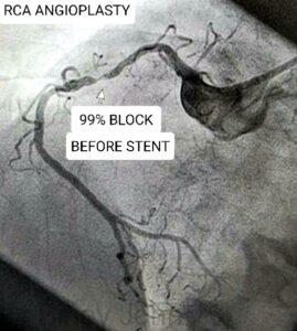
Intra-Vascular Imaging (IVI) is a technology by which images can be obtained from inside the coronary artery by the use of light (Optical Coherence Tomography) or ultra-sound (Intra-Vascular Ultra-Sound).Conventionally, blocks in arteries of the heart are diagnosed with a coronary angiogram, by injecting dye into the arteries of the heart and visualizing them on live x-ray. (Image shows a block as seen by a conventional angiogram using dye)
Subsequently, angioplasty and stenting of the block arealso performed with the guidance of the dye and x-ray technology. However, this has changed for the better, with the advent of IVI.
With the availability of IVI, it is being widely used in cardiology and coronary angioplasty as it helps in viewing the inside of the arteries and obtaininglot of more precise and clearer information which helps in better diagnosis and giving better quality results during angioplasty.
What is Optical coherence tomography?
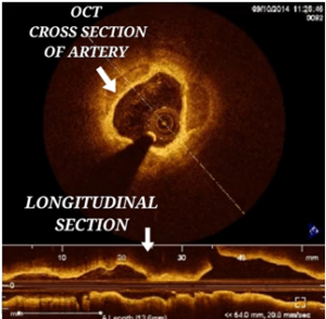
Optical coherence tomography is a technology by which near-infra-red light is emitted through a very thin fibre-optic catheter, which is passed inside the coronary artery. This light helps in obtaining very clear high resolution images from inside the artery. (See image)
How is Optical Coherence Tomography different from Intra-Vascular Ultra-Sound?
The key differences between OCT and IVUS is that the former uses near-infrared light to generate images while the latter uses ultrasound waves to generate images. Besides this, OCT requires injection of dye into the artery to clear the blood from the artery, when the images are being acquired while IVUS does not need this. This makes IVUS the imaging of choice in patients with kidney function impairment as the amount of dye that can be used in kidney failure patients needs to be limited.
In contrast to conventional angiography, OCT helps to obtain very clear anatomical information like the exact diameter of the artery and percentage of the block and thus helps in more accurate selection of the length and diameter of the stent needed for that artery. The measurements by a conventional angiogram, which is only a lumenogram, are often only an approximate and the measurements may even be under-estimated. (See image)
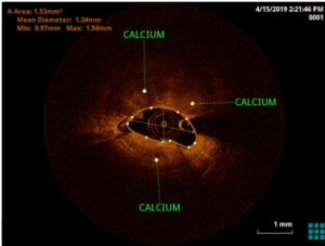
OCT also gives more precise information on the types of the blocks like the presence and percentage of fat, fibrous tissue and calcium in the blocks. Different types of blocks will need different types of balloons and hardware for successful opening of the blocks. This informationfrom OCT aids in selecting the appropriate balloons and hardware to be used in the angioplasty. Thus depending on the morphology, plain balloons can be selected for soft fatty tissue, cutting balloons with blades for fibrous tissue and drills like the rotablator or shock waves or intra-vascular lithotripsy or orbital atherectomy for breaking and opening calcified or rock like blocks. (See image)
Use of OCT after stenting:
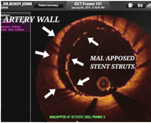
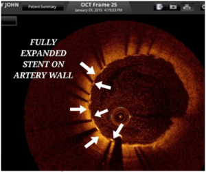
OCT can also be used to see the inside of the stents after angioplasty and to see whether the stents are well expanded. Poorly expanded or mal-apposed stents (See image) can cause complications like clot formation inside the stent and a resultant heart attack.
Such poor expansion of the stent when identified by OCT can be corrected immediately with the use of high pressure balloons. (See image and video)
Watch Video: How does the coronary artery look by OCT after angioplasty and stenting?
Use of OCT in treating blocks which occur inside stents: OCT also aids in treating blocks occurring inside the stents called In-Stent Re-stenosis (ISR), by providing information about the character of the block and also the possible mechanism why the block occurred thus enabling to select the best and ideal hard-ware and techniques to treat the ISR. (See video)
Video: OCT being used to inside a stent to treat In-Stent Restenosis (ISR).
Use of OCT in heart attacks:
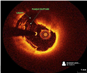
OCT is also useful in understanding the mechanism of heart attacks. Some heart attacks can be caused without any significant blocks, by a mechanism called plaque rupture. Clots are responsible for such heart attacks. (See images and video) Some of these patients may not need stents. OCT aids in making decisions in such patients on the need for stent versus only medical management.

Video: What happens during a heart attack, as seen with OCT imaging?
Imaging with OCT thus aids in getting a lot of information which helps in a clear understanding of blocks and selecting the best hard-ware and ensuring the best results.
Expertise: Dr Binoy John is one amongst the first cardiologists to start using OCT in India, as early as 2014. He has written chapters on OCT and delivered several lectures on use of OCT in angioplasty in national and international conferences.


