
What is the Aortic valve?
Aortic valve is one of the four valves of the heart. It is the valve located between the left ventricle and aorta. It controls forward flow of blood from the left ventricle, which is the pumping chamber of the heart, into the largest artery of the body which is the aorta. It is through the various branches of the aorta that the entire human body gets blood, oxygen and nutrition. The main function of the aortic valve is to prevent backward flow from the Aorta into the left ventricle. The aortic valve normally has three cusps or leaflets.
What are Aortic stenosis and Aortic regurgitation?
When the Aortic valve gets thickened, its opening can get restricted and narrowed, offering resistance to flow and volume of blood pumped from the left ventricle to the aorta. This condition is called Aortic Stenosis. The thickened and restricted aortic valve can also cause leak from aorta into the left ventricle which is called Aortic regurgitation. Aortic stenosis and regurgitation can occur due to many causes. The commonest causes of aortic stenosis are age related or degenerative and Bicuspid aortic valve. Bicuspid aortic valve is a condition where at birth, the person has only two leaflets, causing an injury to the leaflets by the high pressures in the left ventricle and aorta, leading to rapid development of aortic stenosis and or aortic regurgitation.
What are the treatment options for Aortic stenosis and aortic regurgitation?
The treatment options for either can be Surgical Aortic Valve Replacement (SAVR) where the chest and heart are opened surgically and the valve is replaced with a new valve which can be a Mechanical valve (metal) or Tissue valve (Non-metal). This has been the standard treatment.
However, with the evolution of angioplasty, percutaneous techniques have evolved, whereby the Aortic valve can currently be replaced, without opening the chest or heart, under local anaesthesia or deep sedation and sometimes under general anaesthesia, depending on the criticality of the patient. This percutaneous technique is called Trans-catheter Aortic Valve Implantation (TAVI).
The ideal indication for TAVI is a high surgical mortality risk patient. Almost all TAVI is performed for Aortic stenosis and only rarely for aortic regurgitation.
How is Trans-catheter Aortic Valve Implantation performed?
Types of valves used: There are two types of valves which are commonly used. They are the Balloon expandable valves (Sapien series by Edwards) and the Self expanding valves (Evolut series by Medtronic). The technology of the device and the steps ofimplantation of either valves are different.
The Balloon expandable valves are mounted on a balloon and once they are positioned across the Aortic valve, the balloon is inflated and the valve gets implanted over the native aortic valve.
The Self expanding valves are compressed inside a catheter, and once the valve is positioned across the aortic valve, the catheter is removed carefully, exposing the new valve and the valve expands by itself when exposed to the temperature of the blood. On complete removal of the catheter is done, the entire valve gets exposed and gets deployed and starts functioning.
Access: TAVI can be performed from different sites as the femoral artery in the leg, the axillary artery in the chest, the carotid artery in the neck and various other un-conventional routes. The commonest accesses are the femoral artery and the axillary artery.
TAVR with self-expanding valve
Procedure:
The procedure involves a wire being placed inside the left ventricle. It is on this wire that the valve is advanced and positioned across the narrowed and diseased aortic valve. A balloon dilatation of this narrowed valve is done in certain cases.
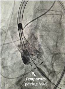
A temporary pacing wire is also positioned in the Right ventricle to pace the heart when balloon dilatation is performed and also when the new valve is being positioned and implanted, as there can be a slowing of heart rate and risk of cardiac arrest during these steps. (See image)
Once the new prosthetic aortic valve is positioned across the aortic valve, the sheath inside which the valve is compressed and kept is released. As the sheath is released, the new prosthetic aortic valve opens up. (See image)
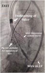
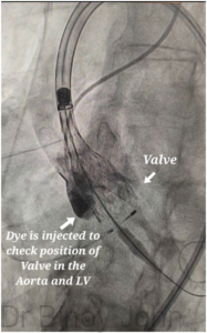
A pig tail catheter placed near the valve is used to inject dye and see if the valve position is perfect. (See image)
Once the position of the valve is confirmed as good, the valve is completely un-sheathed and the valve gets completely released and starts functioning normally. (See image)
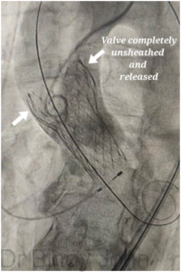
The procedure ends after checking if the valve is opening well, the pressure gradient across the valve and if there are any significant leaks through the sides (para-valvar leak). Repeat balloon dilatation is done if there are significant pressure gradients or if there is any significant para-valvar leak.
Expertise: Dr Binoy John is experienced in TAVR and has trained in GOVKI, Budapest & Cedars-Sinai Heart Institute.
Watch: TAVR with self expanding valves:


